|  
                                 
                                 Abdominal wall - large ventral 
                                  hernias and incisional hernia:  
                                  (Recent developments - the use of imaging and 
                                  Botox injection) 
                                 
                                Morry Brygel 
                                    
                                  Correspondence: 
                                  Professor Brygel 
                                  Melbourne Hernia Clinic 
                                  Melbourne 
                                  Australia 
                                  Email: mbrygel@netspace.net.au; 
                                  www.hernia.net.au  
                                   
                                  Introduction 
                                  Both ultrasound 
                                  and CT scanning have become increasingly useful 
                                  in the diagnosis and management of conditions 
                                  of the abdominal wall, particularly hernias. 
                                  Large ventral, incisional and recurrent hernias 
                                  remain a significant challenge despite the introduction 
                                  of mesh, component separation and laparoscopy. 
                                   
                                  Recurrence rates remain between 7% and 48%. 
                                   
                                  Abdominal wall masses may include subcutaneous 
                                  lipomas or rarely, muscular tumours. Ultrasound 
                                  may also diagnose ruptures of the rectus muscle 
                                  from either a direct blow or excess effort or 
                                  straining. 
                                Hernias 
                                   
                                  Imaging 
                                 
                                Both ultrasound and CT are used in the diagnosis 
                                  and management of hernias. 
                                An ultrasound can demonstrate small Spigelian 
                                  hernias which are often concealed. These can 
                                  even be confused with an inguinal hernia. They 
                                  are not as useful for very large hernias as 
                                  it is difficult to outline the whole defect. 
                                   
                                  The ultrasound can be used in the assessment 
                                  of umbilical and epigastric hernias. The size 
                                  of the defect may be relevant in deciding whether 
                                  a mesh is used, thus facilitating an informed 
                                  consent discussion regarding the use of mesh. 
                                  It may also detect additional small epigastric 
                                  hernias ensuring that they are not missed at 
                                  surgery. An emerging use of ultrasound is as 
                                  a guide to the placement of Botox, in the management 
                                  of large ventral or incisional hernias. 


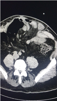
                                  Incisional hernia coronal view 


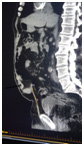
                                  This is a ct sagittal view demonstrating 
                                  a large incisional hernia extending almost to 
                                  the symphyses pubis.  
                                 
                                CT SCANS 
                                  CT scans are most useful for very large ventral 
                                  or incisional hernias, as they accurately locate 
                                  and measure the number and size of the defects. 
                                  This is not feasible with ultrasound alone. 
                                  It is useful for the surgeon to pre-operatively 
                                  anticipate the size of the mesh required and 
                                  the complexity of the operation. 
                                The CT also defines the contents of the hernia, 
                                  be it omentum, bowel or fluid. It may also be 
                                  used to exclude other intra abdominal coexistent 
                                  pathology. 
                                Botox 
                                  A recent important innovation is the use of 
                                  Botox in the management of large ventral hernias. 
                                  Botox is injected under ultrasound control into 
                                  the bellies of the three lateral abdominal wall 
                                  muscles on each side, two weeks prior to surgery. 
                                  The Botox relaxes these muscles and enables 
                                  apposition of the rectus muscles more easily. 
                                  The loss of domain which can impair respiration 
                                  once the hernia is closed is also minimized 
                                  because of the flaccidity of the lateral abdominal 
                                  wall muscles from the Botox. This lasts for 
                                  about 6 weeks. This flaccidity also reduces 
                                  the risk of recurrence.  
                                It also reduces the levels of post operative 
                                  pain which is often significant even with laparoscopic 
                                  repair. The effect lasts for a many weeks further 
                                  reducing the risk of recurrence 
                                In Botox research the scan shows the 3 lateral 
                                  abdominus muscles are lengthened considerably 
                                  . This reduces the size of the defect to be 
                                  closed.  
                                   
                                  The Melbourne Hernia 
                                  Clinic has an educational site devoted to hernias 
                                  and office surgery. A/Prof Maurice Brygel also 
                                  conducts skills workshops for GPS - for details: 
                                  visit www.hernia.net.au 


                                 |